Mada za sehemu hiiCoordinationMada 8
There are numerous types of sensory receptors in the skin which shows some receptors in place.
These receptors are concerned with at least five different senses namely touch, pressure, heat, cold and pain. Some of the skin receptors particularly those concerned with pain are simply the unmyelinated terminal branches of neurons.
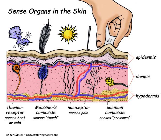
Others are nets of nerve fibres surrounding the bases of hairs. These are particularly important in the sense of touch.
These are stimulated by the slightest displacement of the tiny hairs present on most parts of the body. Other skin receptors are more complex consisting of nerve endings surrounded by a capsule of a specialized connective tissue cells.
NB:
The relative abundance of the various types of the receptors differs greatly e.g. pain receptors are nearly 27 times more abundant than cold receptors and cold receptors are nearly 10 times more abundant than heat receptors.
The receptors are not evenly distributed over the entire body e.g. touch receptors are much more numerous the finger tips than in the skin of the back.
These are widely distributed over the internal body and function primarily in receiving information about the condition of the body itself.
Though the senses receptors mediate are not included in traditional classification of the five, they are of immense importance in the life of an organism. Examples are the stretch receptors (proprioreceptors) in the muscles and tendons, which are involved in the knee jerk reflex.
They are sensitive to the changing tension of muscles and tendons and send impulses to the central nervous system informing it of the position and movements of the various parts of the body
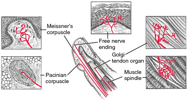
The terminal branches of sensory nerve fibre are intimately associated with several specialized muscle fibre that form an apparatus called neuromuscular spindle.
Other dispersed receptors include those of visceral senses located in the internal organs e.g. receptor in the carotid artery sensitive to carbon dioxide concentration in the blood and to blood pressure.
The firing of such visceral receptors seldom results in sensations (i.e. we are not aware of their action) the responses to the stimulation of visceral receptors produces conscious sense such as thirst, hunger and nausea.
The receptor cells for taste are located in taste buds on the upper surface of the tongue and to a lesser extent on the surface of the pharynx and larynx.
The receptor cells themselves are not neurons but specialized cells with microvillus on their outer ends.
The end of nerve fibres lie very close to these receptor cells, and when a receptor cell is stimulated, it generates impulses in the fibres.

The diagram above shows the structure of the taste bud. Each taste bud contains specialized receptor cells bearing sensory microvilli that are exposed in pits on the tongue surface. The ends of sensory neurons (coloured) are closely associated with these receptor cells.
Adaptations
Adaptation is the decline in the frequency of impulses when a strong or constant stimulus is perceived by a sensory receptor cell e.g. on entering a room you may immediately notice a clock ticking but after a while you become unaware of its presence.
The rate and extent of adaptation in a receptor cell is related to its function and there are types; rapidly and slowly adapting receptors.
Slowly adapting receptors (tonic receptors) register constant stimulus with a slowly decaying frequency of impulses.
The adaptation is thought to be due to decrease in the permeability of the receptor membrane to ions due to sustained stimulation. This progressively reduces the size and the duration of the generator potential and when this falls below threshold level, the sensory neuron ceases to fire the impulse.
Advantage of adaptations
It provides animals with precise information about change in the environment.
- At other times the cells do not send signals thus preventing over loading of the central nervous system with irrelevant and unmanageable information.
- It ensures efficiency and economy of the nervous system.
Rapidly adapting receptor (phasic receptor)
Respond to changes in stimulus level by producing a high frequency of impulses at the movements when the stimulus switched "on" or "off"
E.g. Pacinian corpuscle and other receptors concerned with touch and the detection of sudden changes acts in this way.

This means that while the generator potential from an individual receptor cell may be insufficient to set up an action potential across the synapse. The generator potential from receptor cells may add together or summate and trigger an action potential. This is known as convergence and is a useful adaptation for increasing the sensitivity of a sensory system to low level stimuli.
The role of iris
Iris is a circular sheet of muscles dividing the eye into two chambers.
The pigment it contains gives the eye its colour. The reflex contraction and relaxation of the muscles of the iris control the amount of light entering the eye.
When we look at something our eyeballs are moved in their sockets by muscles so that the pupil at the centre of the iris is pointing at the object of our interest.
Light from the object enters the eye through the pupil and the amount of light entering is controlled by the size of the opening. This in turn is controlled by the Iris muscles.
Accommodation
Accommodation is the reflex mechanism by which light rays from an object are brought to focus on the retina.
It involves two processes which are,
- Reflex adjustment of the pupil size
- Refraction of light rays
Reflex adjustment of pupil size
In bright light
The iris reduces the size of pupil by contracting its circular muscles and relaxing its radial muscles to prevent damage to the light sensitive cells by strong light.

In dim light /poor light intensity
The circular muscles relax and the radial muscle contract opening the pupil aperture as wide as possible to allow the maximum amount of light to ensure the best possible vision.

Changing the shape of the lens
The ciliary muscles are arranged circularly around the ciliary body, the effects of their contractions and relaxations are relayed to the lens by the suspensory ligaments. The lens itself is elastic and its unstretched shape is relatively short and fat.
When ciliary muscles relax, the gap around the lens gets longer, increasing the tension in suspensory ligaments. These in turn pull on the lens making it long and thin. Its ability to bend light is now minimum and it is said to be unaccommodated.
When ciliary muscles contract they reduce the gap around the lens. This reduces the tension in the suspensory ligaments allowing the lens to become short and fat. In this state it is fully accommodated and its ability to bend the light is maximum.
Refraction of light rays
Light rays from a distant object are parallel when they strike the eye. Light rays from a near object are diverging when they strike the eye. Both cases light rays must be refracted or bent to focus on the retina and refraction must be greater for light from near objects.
Refraction occurs when light passes from one medium into another with a different refractive index, and this occurs at the air to the cornea at the surface of the lens.
The degree of refraction at the cornea surface depends on the angle at which light strikes the cornea; also depend upon the distance of object from the cornea.
Most of the refraction occurs in the cornea and consequently the function of the lens is to produce the final refraction that brings light to sharp focus on the retina
The light entering the eye is refracted by its passage through the conjunctiva, cornea, aqueous humour and vitreous humour in exactly the same way regardless of whether it is from a near or a distant object. But by changing the shape of the lens the degree of bending of the light can be altered. Light from distant objects needs relatively little bending to bring it into focus on the retina and so the lens has to be thin. (Because they are almost parallel and not diverging like for near object).
i. Light from a distant object and Light from near object

The role of retina
Retina is a layer of light sensitive cells (i.e. rods and cones and the neurones leading from these photoreceptors to the optic nerve.
The light from the objects is focused into the retina. The retina must then perceive that light and inform the brain of its presence.
In order to do this the retina contains about a hundred million light sensitive (photoreceptors) along with the neurones with which they synapse.
There are two main types of photoreceptors in the retina known as the rods and the cones; shown in the figure below
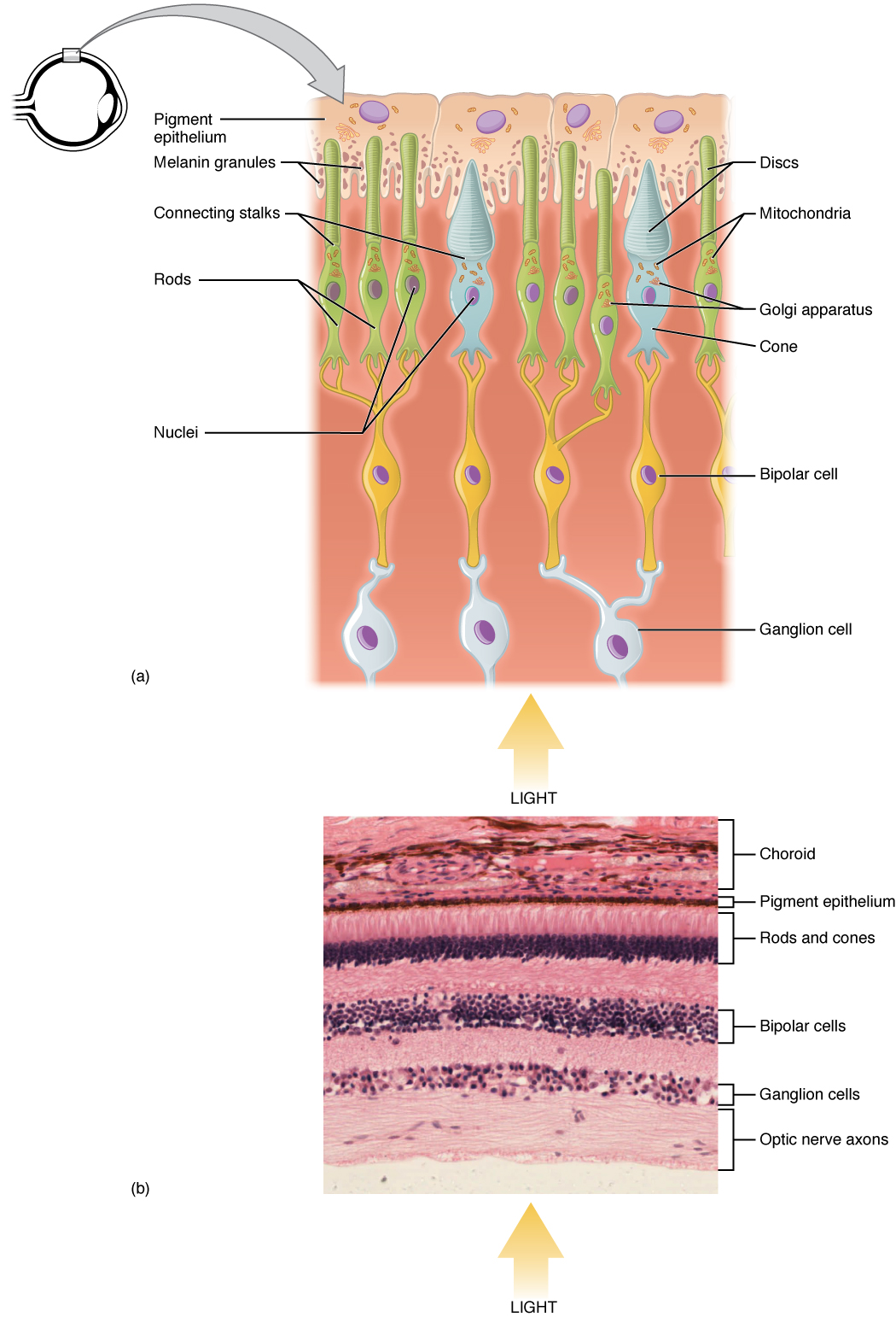
The structure of the retina
The retina is composed of three layers of cells each containing a characteristic type of cell; these are:
- Photoreceptor layer (outermost layer) containing photosensitive cells; the rods and cones partially embedded in the pigmented epithelial cells of the choroid.
- Intermediate layer containing bipolar neurons with synapse connecting the photoreceptor layer to the cells of the third layer. Cells called horizontal and amacrine cells found in this layer enable lateral inhibition to occur.
- Internal surface layer containing ganglion cells with dendrite in contact with bipolar neurons and axons of the optic nerve.
The reason for this somewhat unexpected arrangement is the origin of the retinal cells in the embryo and the way in which the eye is formed during the embryonic development. To add to this confusion; the optic nerve carrying the visual information cross over on their way to the visual cortex in the brain so that the information seen with the right eye is taken to the left side of the brain for processing.
A diagrammatic section through the retina of the eye showing the ultra structure of a rod and a cone.
Outer segments
Outer segment is the light sensitive region where the light acts as a stimulus to the production of generator potential. They contain flattened membranous vesicles filled with photosensitive pigments.
Constriction:
Constriction is a very narrow region between the outer and inner segment.
- It contains two cilia with unusual structure.
- They have inner strands of their 9 + 2 structure missing.
Inner segment
This is packed with mitochondria which produce energy for various processes and ribosomes which synthesize proteins for the vesicles and visual pigments.
Synaptic region.
The cells form synapses with the bipolar cells and several rods synapse with one bipolar cell join to give increased sensitivity to light (convergence phenomenon).
Once connected with one bipolar cell giving great visual activity.
Visual acuity:
is the ability of the eye to resolve two or more stimuli spatially separated. The cells of the human retina
The human retina has the following cells.
- The receptor cells (rods and cones) making the outer layer.
- The bipolar cells which synapse at their tips with receptor cells these make up the in middle layer of retina.
- Ganglion cells: Which synapse at their tip with the bipolar cells; make the third layer. Their axons form the optic nerve, which run from the eyes to the brain.
Roles of horizontal cells and amacrine cells.
- Horizontal cells Synapse with several bipolar neurons; this increases visual acuity and sensitivity of the vision. By exerting lateral inhibition. If they receive stimuli from two rods of equal intensity they cancel out (inhibit the stimuli). They therefore enhance contrast between areas that are strongly stimulated and areas that are weakly stimulated. This makes features such as edges of objects stand out more clearly.
- Amacrine cells (are stimulated by bipolar neurons and synapses with ganglion cells.)
- They transmit information about changes in the level of illumination.
- And hence comment on the mode of action of the nervous system based on the type of neurotransmitter substance they produce.
The rods and cones synapse in the retina with short sensory neurons (bipolar neurons) which themselves synapse in the retina with longer neurons (ganglion cells) whose axons bundled together as the optic nerve, run to the visual centers of the brain.
The presence of several sets of synapses within the retina enables the eye to modify extensively the information transmitted from the receptor cells.
The light sensitivity of rods and cones
Both rods and cones contain light sensitive pigments
In rods the pigment, which is built into the membrane of the flattened vesicles in the outer segment is called rhodopsin
Rhodopsin is made up of a protein scotopsin (opsin) combined with a light absorbing prosthetic group called retinal, which is a derivative of vitamin A.
When a molecule of rhodopsin is struck by a photon of light, the retinal is converted into a slightly different isomer i.e. when converted to trans-isomer; rhodopsin then breaks up into opsin and retinal.
The breaking of rhodopsin sets up a generator potential in the rod and if this is large enough or if several rods are stimulated simultaneously, an action potential is set up in the receptor neuron.
Once bleaching/ breaking of the rhodopsin have occurred, the rod cannot be stimulated again until rhodopsin is resynthesized. It takes energy from ATP produced by the many mitochondria in the inner segment to convert retinal back to the cis-isomer and rejoin with opsin.
Mechanism of photoreception
Rods contain the photosensitive pigment called rhodopsin or visual purple. Rhodopsin is made by combination of a protein called scotopsin with a small light absorbing molecule called retinal (retinene) which is a derivative of vitamin A. In the presence of light rhodopsin decomposes into retinal and scotopsin a process known as bleaching.
Rhodopsin is formed in the absence of further stimulation of light a process known as dark adaptation.
The retinal exists in two isomers. Bleaching leads to the creation of a generator potential in the rod cell which is sufficiently large, to generate an action potential along the neurons leading from the cell to the brain. For daylight cones are used. They contain photosensitive pigments called iodopsin.
Physiology of seeing
The light rays from an object reach the eye and pass through the transparent conjunctiva, cornea, aqueous humour and crystalline lens.
The cornea bends the light rays and the lens causes more bending. The refracted light rays pass through the vitreous humour and finally come to focus at a point on the retina. The point at which the image focuses is called fovea or yellow spot and the image formed is real, smaller than object and inverted.
On the fovea, the light impulses are converted into electrochemical impulses and are sent to the visual area of the brain through the optic nerve. In the brain an interpretation of the size, nature, distance and uprightness of the object is made.
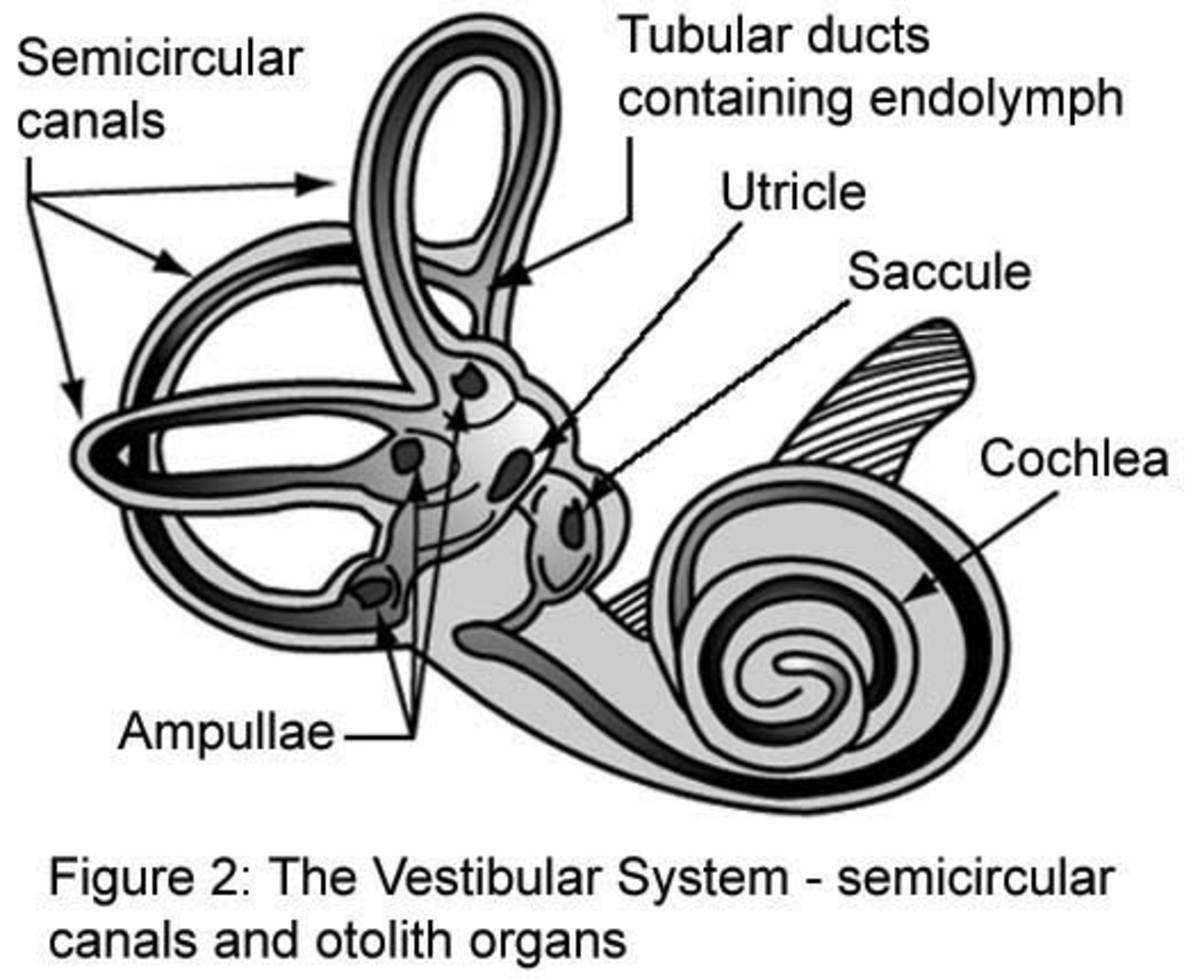
The ear is a sense organ containing mechanoreceptors sensitive to body displacement and sound. Movement and position of the head relative to gravity are detected by the vestibular apparatus composed of semicircular canals, saccule and utricle. All other structures in the ear are involved in receiving amplifying and transducing energy into electrical impulse and promotion of the sensation. The inner ear is principally hearing and balancing part of the ear.

Structure of the membraneous labyrinth
Pointed out earlier, the membranous labyrinth is involved in hearing and balance. It is found in the inner ear. It is structurally composed of three semicircular canals that lie at right angles to one another. The canals arise from a swollen utricle. Below is a high coiled structure that is involved in hearing. The saccule and a connection between the ampulla is known as ductus utriculi.
Cochlea and hearing
Cochlea is a spiral subdivided into three layers vestibular canal and tympanic canal contains perilymph and median canal which contain endolymph. The basilar membrane separates the median and tympanic canals and supports sensory hair cell that can be brought into contact with the tectorial membrane above.
This unit consists of basilar membrane, sensory cells and tectorial membrane is called organ of Corti and is the region where transduction of sound into electrical impulse occurs.

Mechanism of hearing
Sound waves are directed toward the inner ear through the external auditory meatus where they cause the tympanic membrane to vibrate. In the middle ear the vibration of the tympanic membrane are transferred across the oval window by movement of the three ear ossicles, the malleus, incus and stapes.
The vibrations are then transmitted into the inner ear where they cause perilymph of the vestibular canal to vibrate and these are transmitted via Reissner's membrane to the endolymph in the median canal.
From there they are transferred to the basilar membrane and the perilymph in the tympanic canal, and are finally dissipated into the air of the middle ear as vibration of the round window.
Vibration of basilar membrane pushes the sensory hair against the tectorial membrane and forces the two membranes to slide past each other. The distortion produced in the sensory hair cells due to the shearing forces causes a depolarization of the sensory cells, the production of generator potential, and initiation of action potentials in the axons of the auditory nerve. The latter transfer the impulse to auditory part of the brain where an interpretation of the pitch, note, intensity and quality of the sound is made.
The mammalian ear and balance
Several parts of the body are involved in maintaining balance at and during movements. The parts that are involved include eyes, receptors in joints and muscles etc. however vital information about position and movements of the head is provided by the vestibular apparatus of the ear, the utricle, saccule and semicircular canals.
The basic sensory receptor in these structures consist of the hair cells attached to dense structures supported in the endolymph, the region of the walls of utricle and saccule, the maculae contains granules called otoconia in association with receptor cells.
The otoconia responds to gravitational pull and mainly detect the direction of movement of the head with respect to gravity.
The utricle responds to vertical movement of the head e.g. when the body is upside down.
The saccule responds to lateral movement of the head. The semicircular canals respond to rotational movements of the head and they contains cupulae that work in the same way as maculae.
Linear acceleration is detected by both maculae and ampullae.
Adaptation of organ of Corti
- It has vibration and movable membranes
- Sensory cells that detect sound waves
- Has auditory nerve for carrying electrical impulse to the hearing part of the brain
- Has vibratory fluid which amplify sound vibrations.
Mwalimu
Unasoma somo hili? Niulize nikuelezee chochote kilichomo.
Ingia ili kumuuliza Mwalimu wa AI wa Sonza kuhusu mada hii.
Ingia ili kuulizaMajadiliano
Hakuna maswali bado