Mada za sehemu hiiCoordinationMada 8
- The cytoplasm inside the axon, known as the axoplasm, has a high concentration of potassium ions (K⁺) and a low concentration of sodium ions (Na⁺).
- In contrast, the fluid outside the axon has a low concentration of K⁺ and a high concentration of Na⁺.
- A potential difference (charge) exists across the cell surface membrane of all cells. This potential is typically negative inside the cell relative to the outside.
- The potential difference across the membrane when the neuron is at rest is called the resting potential, which is approximately -70 mV. The negative sign indicates that the inside of the cell is more negative than the outside.
Key characteristics of the resting neurone
- Selective Permeability:
- The axon membrane is relatively impermeable to sodium ions (Na⁺).
- It is freely permeable to potassium ions (K⁺).
- Sodium-Potassium Pump:
- The axon membrane contains an active sodium-potassium pump that uses ATP to transport ions.
- It moves sodium ions (Na⁺) out of the axon and potassium ions (K⁺) into the axon.
- This pump reduces the concentration of sodium ions inside the axon. Since Na⁺ is pumped out and cannot diffuse back in, it maintains a low internal Na⁺ concentration.
- At the same time, K⁺ ions are pumped in but can diffuse out along their concentration gradient.
- Polarization:
- As a result of these ion movements, the inside of the cell becomes slightly negatively charged relative to the outside.
- This state is known as polarization, and the potential difference of -70 mV across the membrane is termed the resting potential.

An action potential does not remain confined to a single point on the axon. Instead, it travels along the entire length of the nerve fiber.
How impulses are propagated
- When an action potential is generated at one point, it causes local ion currents.
- These local currents cause depolarization of adjacent areas of the membrane.
- As a result, the action potential is continually generated along the axon in one direction.
- The refractory period prevents the impulse from moving backward.

In myelinated vertebrate nerves, the mechanism of propagation is strongly more complex. Ions can only pass freely into and out of the axon at the nodes of ranvier which are about 1mm apart.
This means that action potential can only occur at the nodes of ranvier and so they appear to jump from one node to the next as the diagram shows. The effects of this is to speed up transmission as the concentration movements associated with the action potential occurs much less frequently taking less time. This condition is known as saltatory condition from the Latin verb which means to jump.

The arrival of an impulse at the synaptic knob increases the permeability at the synaptic membrane to calcium ions.
Calcium ions therefore move into the synaptic knob along concentration gradients the effect of these calcium ions is to cause the synaptic vesicles containing transmitters' substance to move to the pre-synaptic membrane. Each vesicle fuses with the membrane and release the transmitters.
Some of the vesicles fuse with the membrane and release the transmitters. Some of the vesicles fuse with the membrane and release the transmitter substance into the synaptic cleft.
The transmitters diffuse across the gap and become attached to the specific protein receptor sites on the post synaptic membrane.
As a result, ions channels are opened and there is usually a local depolarization and influxmof sodium ions, causing an excitatory post synaptic potential (EPSP) to be set up. If there are sufficient of the potential the positive charge in the postsynaptic cell build up to the threshold level and an action potential in set up this then travels on a long the post synaptic neuron.

Once the transmitter has its effects it is destroyed by enzymes. This is very important because unless the transmitter is removed from the synaptic cleft subsequent impulses would have as effect, as the receptors on the post synaptic membrane would the entire bound.
Nerves have to communicate not only with each other but with receptors and effectors as well. Motor nerves need to communicate with muscles. Where a motor nerve and muscles fibre meet a special kind of synapse is formed known as a neuromuscular junction.
The membrane of the muscle fibre is very folded in this region and forms a structure known as an endplate to which the end of the motor nerve joins. Electron microscope shows us that the structure of the neuro muscular junction is remarkably similar to that of any other synapse as the figure below shows. The end of the motor neurons is full of mitochondria and synaptic vesicles which contain acetylcholine. It appears that when an impulse arrives at the end of the motor neurone acetylcholine is discharged into the synaptic cleft.
As a result of its effect on the postsynaptic membrane an end potential is set up which can be recorded, If sufficient end plate potentials are set up on action potential is fired off on the muscle fibre spreading through the tubules and leading to a contraction of the muscle.


Neurons interact in a variety of complex ways. Sometimes single nerve fibre will carry an action potential to a synapse with another cell and transmission. But in many cases the situation is much more complex than this. Often a single synaptic knob does not release enough transmitter substance to set up an action potential in the post synaptic fibre however, if two or more synaptic knobs are stimulated and release transmitters at sometimes onto the same post synaptic membrane the effects add together and a post synaptic action potential results. This is known as spatial summation; as illustrated below. In other cases, a single knob does not release enough transmitter substance to stimulate the post synaptic nerve fibre, but if a second impulse is received from the same knob in quick succession an action potential results. This effect is known as temporal summation (i.e. adding over time). It involves facilitation in other words, the first impulse does not trigger off a response but it has an effect which make easier (facilitate) the passage of the next impulse. The arrival of the impulse at one synaptic knob triggers an action potential in the post synaptic fibre.
Spatial summation
Action potential needs to arrive at several synapses at once to release the amount of neurotransmitter required to trigger as action potential in the postsynaptic fibre.

Temporal summation
One action potential arises and although it does not release sufficient transmitter substance itself to set up another potential it makes it easier for the next impulses which arises to do so.

Definition; synapse is a region where the branches of an axon are in contact with the dendrites of another neurone.
Types of synapses
Chemical synapses
These are synapses that neurons communicate with each other by means of neurotransmitters.
The neurons are not in direct contact with each other, they join at a synapse which have synaptic cleft, a small gap of about 20nm.
The neurotransmitter is released from one membrane is the synapse the pre-synaptic membrane then it diffuses across the synaptic cleft and binds into receptors on the post synaptic membrane. The post synaptic cell may be in an effectors organ such as muscle or gland.
Types of chemical synapses
The chemical synapses are of two kinds; excitatory synapses and inhibitory synapse.
Excitatory synapse
These are synapses where the pre synaptic neurone releases neurotransmitter that makes the post synaptic membrane more excitable and more likely to generate nerve impulses.
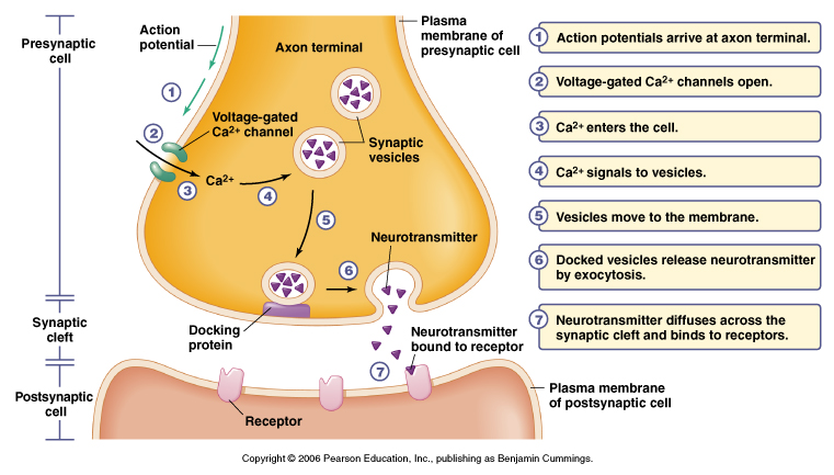
The diagram above shows various events takes place in a chemical synapse which uses acetylcholine as a neurotransmitter.
Acetylcholine is synthesized within the pre synaptic knob and stored in special organelles called synaptic vesicles (some neurotransmitters neither decrease nor increase the tendency of a postsynaptic cell to fire an action potential instead they work like hormones; e.g. noradrenaline released by axons of the sympathetic nervous system has an effect to on cells similar to that of the hormone adrenaline. When an action potential reaches the presynaptic membrane, it depolarizes the membrane, that is; it makes the membrane less negative than at rest. This depolarization triggers the opening of calcium ions channels in the pre synaptic membrane.
The calcium ions diffuse into the presynaptic knob; causing the pre synaptic vesicles containing acetylcholine to migrate and fuse with the presynaptic membrane.
The acetylcholine is released into the synaptic cleft and diffuses across the synapse. The synapse then it binds to specific protein receptor molecules on the post synaptic membrane a process known as receptor activities.
Receptor activation causes sodium ion channels to open making the membrane more permeable and produce a graded potential, if enough acetylcholine is released the graded potential may become large enough an action central.
If an excitatory synapse receives a continuous stream of action potential at high frequency eventually transmission across synapse stops.
This is because the neurotransmitter cannot be resynthesised fast enough and it runs out. The synapse becomes fatigued (i.e. adapted).
Acetylcholine is then broken down by acetylcholinesterace to its constituents groups acetic acid (or ethanoic acid) and choline groups; which diffuses back into the pre synaptic membrane and used to resynthesize the neurotransmitter under the influence of ATP from the mitochondria concentrated here and refilled in vesicles for future transmission.
Inhibitory synapse
These release neurotransmitter that makes the postsynaptic membrane less excitable and less likely to transmit an impulse.
E.g. In mammals, they occur in nerve pathways which central rapid eye movements, they are also common in the heart.

Diagram above: An electrical synapse in which the nerve impulse is transmitted through the protein pores that line the cytoplasm of the two cells
Mwalimu
Unasoma somo hili? Niulize nikuelezee chochote kilichomo.
Ingia ili kumuuliza Mwalimu wa AI wa Sonza kuhusu mada hii.
Ingia ili kuulizaMajadiliano
Hakuna maswali bado