Mada za sehemu hiiGeneticsMada 7
Deoxyribonucleic acid (DNA)
Chemical nature
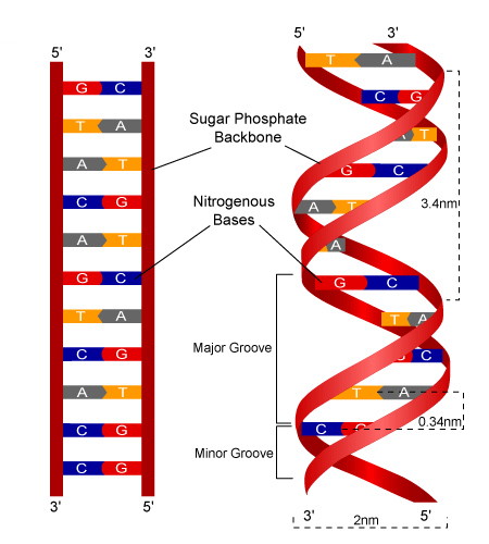
DNA is chemically composed of the following substances:
- Deoxyribose sugar — This is a pentose (5-carbon) sugar.
- Organic or nitrogenous bases — These are of two categories.
- Purine bases — These are Adenine (A) and Guanine (G).
- Pyrimidine bases — These are Cytosine (C) and Thymine (T).
Base pairing rules
- Since DNA is a double stranded molecule, the bases on the two strands appear in pairs being held together by the hydrogen bonds.
- The strands run in opposite directions, that is, they are antiparallel.
- The base pairing rules make the chains complementary.
- According to Watson-Crick model of DNA structure, a purine pairs with a pyrimidine. The rules are that:
- Adenine pairs with thymine and the two bases are held together by two hydrogen bonds.
- Cytosine pairs with guanine and the two bases are held together by three hydrogen bonds.
- Phosphate group — derived from phosphoric acid.
- Protein — over the surface of DNA, there is a histone protein coat.
Chemical bonds
There are two types of chemical bonds.
- Phosphodiester bonds — these hold the nucleotides together.
- Hydrogen bonds — these hold the complementary base parts together.
Diagrammatic structure of DNA
Role of DNA in protein synthesis
This role of DNA is that it instructs the cell of the types of amino acid that should be incorporated to form a protein molecule. That is, the message contains the information about the types of amino acids that should be joined up forming the protein molecules.
- Storing genetic information: DNA carries the instructions for making all proteins in the body. These instructions are stored in the form of genes.
- Providing the template for mRNA: DNA acts as a template during transcription, where a specific gene is copied into messenger RNA (mRNA). This mRNA carries the code for making a protein.
- Determining the sequence of amino acids: The order of bases (A, T, C, G) in the DNA determines the order of amino acids in a protein. Each three bases (called a codon) codes for a specific amino acid.
- Controlling the type of protein made: DNA controls which proteins are made by determining which genes are expressed (transcribed) at a specific time.
- Passing information to future generations: DNA ensures that the instructions for making proteins are passed from one generation of cells to the next during cell division.
- Repairing and maintaining itself: DNA has mechanisms to repair itself to ensure the instructions for protein synthesis remain accurate and error-free.
The features of DNA account for its metabolic stability
- Possession of a histone protein coat.
- The helical nature increases mechanical strength.
- The chemical bonds, i.e., hydrogen and phosphodiester bonds, increase mechanical strength.
Evidence for the role of DNA in inheritance
It took many years to clarify whether the genetic material was DNA or the protein in the chromosomes. It was suspected that protein might be the only molecule with sufficient variety of structure to act as genetic material.
Evidence from bacteria
In the days before development of antibiotics, pneumonia was often a fatal disease. It was intended in developing a vaccine against the bacterium Pneumococcus which was one form of pneumonia.
Two forms of pneumococcus were known, one covered with a gelatinous capsule, one virulent (disease-producing) and the other non-capsulated and non-virulent. The capsule protects the bacterium in some way from attack by the immune system of the host.
Griffith hoped that by injecting the patients with either the non-capsulated or the heat-killed capsulated forms, their bodies would produce antibodies which would give protection against pneumonia. In a series of experiments he injected with both forms of pneumococcus and obtained the results shown in a table below. The dead mice revealed the presence of live capsulated forms in their bodies. On the basis of these results Griffith concluded that something must be passing from the heat-killed capsulated forms to the live non-capsulated forms which caused them to develop a capsule and become virulent.
However, the nature of this transforming principle, as it was known, was not isolated and identified until 1944.
Results of Griffith's experiments

- A representative portion of DNA which is about to undergo replication is shown.
- DNA polymerase causes the two strands of the DNA to separate.
- The DNA polymerase completes the splitting of the strand. Meanwhile, free nucleotides are attracted to their complementary bases.
- Once the nucleotides are lined up, they are joined together. The remaining unwound bases continue to attract these complementary nucleotides.
- Finally, the nucleotides are joined to form a complete polynucleotide chain. In this way, two identical strands of DNA are formed. As each strand retains half of the original DNA material, this method of replication is called semi-conservative method.
The three theories of DNA replication illustrated

Differences between DNA and RNA
| DNA | RNA |
|---|---|
| Double stranded polynucleotide molecule | Single stranded polynucleotide molecule |
| The pentose sugar is deoxyribose | The pentose sugar is ribose |
| The pyrimidine base is thymine | The pyrimidine base is uracil |
| It is found in the nucleus | It is found in the cytoplasm |
| It is constant in the cell | The amount of RNA is variable |
| It is more stable | It is less stable |
| It has high molecular mass | It has low molecular mass |
| The ratio of A to T and G to C is always 1 | The ratio of A to U and G to C is variable |
| Only one basic form, but with an infinity variety within that form | Three basic forms, messenger, transfer and ribosomal RNA |
| It is permanent | It exists temporarily for short periods |
The nature of genes
What are genes?
Mendel defined gene as a unit of inheritance. This is an acceptable definition of gene but it does not tell us anything about the physical nature of gene.
Below are ways of overcoming this objection.
A unit of recombination
It was shown that a gene was the shortest segment of a chromosome which is separated from adjacent segments by crossing over. This definition regards a gene as the specific region of chromosome determining a distinct character in the organism.
A unit of function
- It is known that genes code for proteins;
- Therefore, a gene is the DNA code for a polypeptide.
- Since some proteins are made up of more than one polypeptide chain and are coded by more than one gene.
The genetic code
The genetic code is the relationship between nitrogenous bases on the DNA and amino acids. It was suggested that the genetic information which passed from generation to generation and which controlled the activities of the cell, might be stored in the sequence for the production of protein molecules. It became clear that this sequence in the DNA must be a code for the sequence of amino acids in protein molecules. The relationship between bases and amino acids is known as the genetic code. In other words, the genetic code is a means by which the genetic information in DNA controls the manufacture of specific proteins by the cells. The problems remaining were to demonstrate that a base code existed, to break the code and to determine how the code is translated into the amino acid sequence of a protein molecule.
The code is a triplet code.
There are four bases in the DNA molecules
- Adenine (A)
- Guanine (G)
- Thymine (T)
- Cytosine (C)
Each base is a part of a nucleotide and the nucleotides are arranged as a polynucleotide chain (strand). The sequence of bases indicated by their first letters (alphabets) are responsible for carrying the code that results in the synthesis of potentially infinite number of different protein molecules. There are 20 common amino acids used to make protein and the bases in the DNA must code for them. If one base determined the position of a single amino acid in the primary structure of a protein, the protein could have only four different amino acids. If a combination of two base pairs coded for each amino acid, then 16 amino acids could be specified in the protein molecule. Only a code composed of three bases could incorporate all 20 amino acids into the structure of protein molecules. It was therefore proved that the code is indeed a triplet code, meaning that three bases is the code for one amino acid.
Problems
Using different pairs of the bases A, G, T and C, list the 16 possible combinations of bases that can be produced.
Answer:
| Base | A | G | T | C |
|---|---|---|---|---|
| A | AA | AG | AT | AC |
| G | GA | GG | GT | GC |
| T | TA | TG | TT | TC |
| C | CA | CG | CT | CC |
The mathematical expression is
Where: X = number of bases and Y = number of bases used.
- It is thus a combination of three nitrogenous bases, a three-lettered word of AGC, AUA, GCA, etc.
Features (Characteristics) of the genetic code
- It is a triplet of bases in the polynucleotide chain that codes for an amino acid in the polypeptide chain.
- The genetic code is degenerate, i.e., a given amino acid can be coded for by more than one codon (codons are complementary triplets in the mRNA).
Example:
| Amino acid | Codons |
|---|---|
| Alanine | GCU, GCC, GAC, GCG |
- The genetic code is universal, i.e., the same triplet codes for the same amino acids in all organisms.
- The genetic code can be punctuated, i.e., it has got the 'start' and 'end' signals.
- The genetic code is non-overlapping. For example, if the base sequence is ACAGAGUCGGAC, then this will be read as ACA/GAG/UCG/GAC and not ACA/CAG/AGA.
- The genetic code sequence has got no commas, e.g., AAU, GCG, GAC, etc. This is because the bases are continuously sequenced on the DNA or RNA strand.
Nonsense codons
These codons do not code for amino acids; they primarily mark the end point of polypeptide chains. They act as stop signals for the termination of polypeptide chains during translation.
Protein biosynthesis
'DNA makes RNA and RNA makes Protein'
Protein synthesis is a mechanism by which a protein molecule is constructed by joining the amino acids with the peptide bonds according to the instruction in the mRNA coded from DNA.
Stages of protein synthesis
- Synthesis of amino acids.
- Transcription (formation of mRNA).
- Amino acid activation.
- Translation.
The site for protein synthesis is the ribosome. These proteins synthesized may have a structural role such as keratin and collagen, or a functional role such as insulin, fibrinogen and, most importantly, enzymes which are responsible for controlling all metabolism. It is the particular range of enzymes that determines what type of cell it becomes. This is the way in which DNA controls the activities of a cell. The instructions and information for the manufacture of enzymes and all other proteins are located in the DNA. However, the actual synthesis of protein occurs in the ribosomes in the cytoplasm. Therefore, a mechanism had to exist for carrying the genetic information from the nucleus to the cytoplasm. This link was messenger RNA.
Adaptations of the ribosome to protein synthesis
- Presence of appropriate enzymes that catalyze the synthesis of polypeptide bonds between the amino acids.
- Presence of receptor site for messenger RNA attachment.
- Presence of rRNA for attracting other types of tRNA towards the ribosome.
- Ability to read and 'translate' the message contained in the codes of mRNA.
Mechanism of protein synthesis
There are four main stages in the synthesis of protein:
Synthesis of amino acids
In plants, the formation of amino acids occurs in mitochondria and chloroplasts in a series of stages:
- Absorption of nitrates from the soil.
- Reduction of those nitrates to the amino group (NH).
- Combination of those amino groups with a carbohydrate skeleton (e.g., alpha-ketoglutarate from Krebs cycle).
- Transfer of the amino group from one carbohydrate skeleton to another by a process called transamination.
Animals usually obtain their amino acids from the food they ingest, although they have capacity to synthesize their own non-essential amino acids.
Transcription (formation of mRNA)
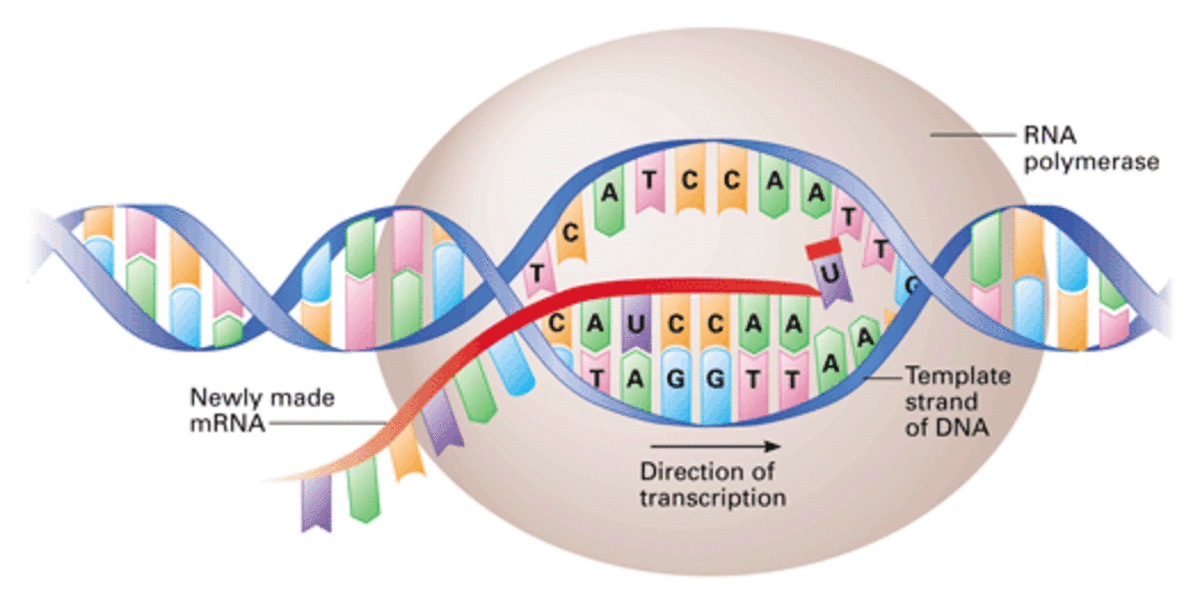
- This is a mechanism by which the base sequence of a section of DNA representing a gene is converted into a complementary base sequence of mRNA.
- In this process, a complementary mRNA copy is made from a specific region of the molecule which codes for a polypeptide.
Mechanism of transcription
A specific region of the DNA molecule, called a cistron, unwinds. This unwinding is a result of hydrogen bonds between base pairs in the DNA double helix being broken. This exposes the bases along each strand and one of these strands is selected as a template against which mRNA is constructed. This mRNA molecule is formed by linking free nucleotides under the influence of RNA polymerase and according to the rules of base pairing between DNA and RNA.
Table to show the RNA bases which are complementary to those of DNA
| DNA bases | Complementary RNA bases |
|---|---|
| A (Adenine) | U (Uracil) |
| G (Guanine) | C (Cytosine) |
| T (Thymine) | A (Adenine) |
| C (Cytosine) | G (Guanine) |
When the mRNA molecule has been synthesized, it leaves the nucleus via the nuclear pore and carries the genetic code to the ribosomes. Along the mRNA is a sequence of triplet codes which have been determined by the DNA. Each triple is called a codon. When sufficient numbers of mRNA molecules have been formed from the gene, the RNA polymerase molecule leaves the DNA and the two strands 'zip up' reforming the double helix.
Amino acid activation
Activation is the process by which amino acids combine with tRNA using energy from ATP. Each type of tRNA binds with the specific amino acid which means there must be at least 20 types of tRNA. Each type differs, among other things, in the composition of a triplet of bases called anticodons which terminate in the CCA. It is to the free end that the individual amino acid is attached. The tRNA molecules with attached amino acids form an amino acid-tRNA complex known as aminoacyl-tRNA and their formation is under the enzyme aminoacyl-tRNA synthetase. The combination now moves towards the ribosome.

Translation
- Translation is the mechanism by which the sequence of bases in an mRNA molecule is converted into a sequence of amino acids in a polypeptide chain.
- Several ribosomes may become attached to a molecule of mRNA like beads on a string and a whole structure is known as a polyribosome or polysome.
- The advantage of such an arrangement is that it allows several polypeptides to be synthesized at the same time.
- The first two mRNA codons (a total of 6 bases) enter the ribosome. The first codon binds the aminoacyl-tRNA molecule having the complementary anticodon and which is carrying the first amino acid (usually methionine) of the polypeptide being synthesized.
- The second codon then also attracts an aminoacyl-tRNA molecule showing the complementary anticodon.
- The function of the ribosome is to hold in position the mRNA, tRNA and the associated enzymes controlling the process until a peptide bond forms between the adjacent amino acids.
Once the new amino acid has been added to the growing polypeptide chain, the ribosome moves one codon along the mRNA. The tRNA molecule which was previously attached to the polypeptide chain now leaves the ribosome and passes back to the cytoplasm to be reconverted into a new aminoacyl-tRNA molecule.
This sequence of the ribosome 'reading' and 'translating' the mRNA code continues until it comes to a codon signaling 'stop'. These terminating codons are UAA, UAG and UGA. At this point, the polypeptide chain, now with its primary structure as determined by DNA, leaves the ribosome and translation is complete.
The main steps involved in translation may be summarized under the following headings
- Binding of mRNA to ribosome.
- Amino acid activation and attachment to tRNA.
- Polypeptide chain initiation.
- Chain elongation.
- Chain termination.
- Fate of mRNA.
The polypeptides so formed must now be assembled into proteins. This may involve the spiraling of the polypeptides to give a secondary structure, its folding to give a tertiary structure and its combination with other polypeptides and/or prosthetic group to give a quaternary structure. If the ribosome is attached to ER (rough ER), the protein enters the ER to be transported.
Introns and exons
It was discovered that the DNA of a eukaryotic gene is longer than its corresponding mRNA. It should be the same length because the messenger RNA is a direct copy. It was discovered that immediately after the mRNA is made, certain sections of the molecule are cut out before it is used in translation. The sections of the gene that code for the unused pieces of RNA are called introns. The remaining sections of the gene that code for the protein are called exons.
Mwalimu
Unasoma somo hili? Niulize nikuelezee chochote kilichomo.
Ingia ili kumuuliza Mwalimu wa AI wa Sonza kuhusu mada hii.
Ingia ili kuulizaMajadiliano
Hakuna maswali bado