Mada za sehemu hiiCytologyMada 9
- Concept of cytology
- Cell Theory
- Cell structure and function
- Cell Differentiation
- Carbohydrates
- Lipids
- Proteins
- Enzymes
- ATP
Structure of cells and their functions
What is a cell?
- A cell is the basic structural and functional unit of life.
- It is a tiny, 3D sac-like structure filled with cytoplasm (a jelly-like fluid), in which various organelles are suspended.
- The cell is surrounded by a membrane, and in plant cells, there is also a cell wall made of cellulose for extra support.
Ultra-structure of the cell
The detailed structure of the cell (organelles) is called the ultrastructure.
It is studied using:
- Electron microscope
- Cell fractionation (a lab method to separate organelles)
Functions within the cell
All metabolic and physiological activities (like respiration, protein synthesis, etc.) take place inside the cell.
Cell theory highlights
- All living organisms are made of one or more cells.
- The cell is the basic unit of life.
- New cells arise from pre-existing cells through cell division.
- The origin of the first cell is not explained by this theory.
Exception – Viruses
- Viruses are not made of cells and are considered non-living outside a host.
- However, inside a host, viruses behave like living organisms (they reproduce and carry out limited functions), challenging the traditional cell theory.
Types of cells
Prokaryotic cells
General characteristics
- They lack well organised nuclei. That is, their nucleus has no nuclear membrane, therefore, the nuclear materials are freely suspended in the cytoplasm.
- They have small, circular and naked DNA. That is, the DNA is not associated with histone protein coat to form chromosomes.
- They have few and small ribosomes of 70s sedimentation coefficient.
- They lack membrane bound organelles such as mitochondria, Golgi bodies, and plastids.
- The cell wall is chemically composed of a carbohydrate-protein complex called peptidoglycan or murein.
- Cilia and flagella, if present do not arise from basal bodies, and they lack microtubules of a "9+2" arrangement pattern.
Structure of prokaryotic cells
The prokaryotic cell does not have a defined nucleus, as it lacks a nuclear membrane. Almost all prokaryotes have a protective cell wall that prevents them from bursting in hypotonic conditions. Such cell walls have different components depending on the type of organism.

Functions of parts of prokaryotic cells
- The cell wall of prokaryotic cells provides structural and protective functions. In some prokaryotes, the cell wall is surrounded by a thin sheath, while in others, it is surrounded by a slim capsule.
- The cytoplasm of prokaryotes is surrounded by a single cell membrane, and all metabolic processes such as protein synthesis, respiration, and replication take place within the cytoplasm.
- Genetic material in the form of a single circular DNA located in the specified region within the cytoplasm. This controls hereditary functions of the cell.
- The flagellum found in most aquatic and soil bacteria aids in movement by a unique process of spinning on an axis like a propeller.
- The pili, a structure located on the cell wall help bacteria to increase the chances of survival by undergoing conjugation or by enabling anchoring to its host or substrate.
- The mesosomes which are infoldings of the cell surface membrane which act as sites of respiratory enzymes.
- Granules of food stores are used as respiratory substrates.
Eukaryotic cells
These cells are characterised by the following features:
- They are relatively large in size, ranging from microscopic to macroscopic.
- They have true or well organized nuclei with nuclear membranes.
- They have large and numerous ribosomes with the sedimentation speed of 80s (the 's' stands for the name Svedberg, but is also a unit of measurement).
- They have membrane bound organelles, such as mitochondria and plastids.
- The cell walls, if present, are chemically composed of cellulose and/or chitin.
- They have large, helical DNA which is associated with histone protein to form chromosomes.
- Cilia and flagella, if present, arise from basal bodies and contain microtubules that are arranged in a "9+2" pattern.
The cells of eukaryotic have three basic parts
- The plasma membrane.
- The cytoplasm.
- The nucleus.
Plasma membrane
-
This is also called the cell surface membrane, plasma membrane, or plasma lemma.
-
It separates the contents of the cells from the external environment, controlling the exchange of materials.
-
In animal cells, it is the outermost layer, whereas in plant cells, it is located beneath the cell wall.
- E.g. Neurilemma in neurons
- Muscle cells – sarcolemma
Structure of the cell membrane
There are two models suggested by different scientists to describe the cell membranes:
Daniel-Davson model
-
According to Daniel and Davson, the membrane is structurally composed of two chemical substances that form their own layer:
-
Protein layer made up of molecules.
- The layer is continuous and lacks pores.
-
Phospholipids (at least two layers) oriented with:
- Polar (hydrophilic) ends near the surface
- Non-polar (hydrophobic) hydrocarbon chains in the interior of the membrane, as far as possible from the surrounding water.
-
-
According to this model, the membrane is structurally rigid, static, and non-dynamic.

Strength of the model
- The model suggests that the membrane is composed of proteins and lipids. Amphipathic (double) nature of phospholipids such as phospholipids molecule has a polar head (hydrophilic) and a non polar tail (hydrophobic).
Weakness of the model
- The model suggests that the protein layer is continuous. Researches done by scientists show that the protein layer is discontinuous.
- The membrane is static is a wrong concept since the membrane is a dynamic ever changing structure.
- Lack of pores in protein layers.
- The protein molecules in a membrane have pores for passage of materials.
- The model does not indicate the presence of a carbohydrate.
The fluid mosaic model
-
The model was put forward by Singer and Nicolson (1972) in order to modify the Daniel and Davson model.
-
According to the fluid mosaic model:
- The membrane is an ever-changing structure in which the mosaic protein floats on the lipid bilayer, acting as a fluid.
- Proteins in this model do not form a continuous layer covering both sides of the membrane, as proposed by the Daniel and Davson model.

According to this model, the membrane has 3 constituents:
- Lipids (45%)
- Proteins (45%)
- Carbohydrates (10%)
Lipids
There are two types of lipids:
-
Glycolipids
- These are lipids associated with short carbohydrate chains.
-
Phospholipids
-
These are lipids associated with phosphates.
-
They form 2 layers, i.e., the phospholipid bilayer.
-
Each phospholipid consists of:
- A polar head (hydrophilic)
- A non-polar tail (hydrophobic)
-
Act as a fluid and move about rapidly in their own layer.
-
Since phospholipids are constantly in motion, the membrane is described as being fluidly.
-
Roles of phospholipids
- Form the basic structure of the membrane.
- Determine the fluidity of the membrane.
- Allow the passage of fat soluble substances.
NB: cholesterol is a type of steroid located in between phospholipids keeping them fluidly.
Roles of cholesterol
- Disturb the close package of phospholipids, keeping them fluid.
- Increase the flexibility of the membranes by allowing relative movements of the bilayers without actual displacement, because it acts as an unsaturated fatty acid lubricating bilayer.
Proteins
-
These exist as globular in the membrane, i.e., they never form a continuous layer.
-
Within protein molecules or between adjacent ones, there are poles, which may be:
- Hydrophobic
- Hydrophilic
-
Since phospholipids are always in constant motion (fluid), proteins float in it, forming a fluid mosaic model.
-
The proteins are organized in a particular pattern known as a mosaic.
-
There are protein molecules that:
- Extend/transverse both layers of membranes
- Partially embedded in the membrane – these are called intrinsic proteins
-
Some proteins float freely inside the membrane – these are called peripheral or extrinsic proteins
Enzymes
Catalyze different metabolic reactions
Receptor molecule
-
Some act as receptors for chemical stimuli
- Example: Hormones
Identity markers
- These are glycoproteins
- They have different shapes in every kind of a cell
- They have specific side chains, thus are recognized by other cells and behave in an organized manner
Energy transfer
-
In some physiological processes such as photosynthesis and respiration, some proteins are involved in energy transfer
-
These are found in special forms of membrane in:
- Chloroplasts
- Mitochondria
Carbohydrates
These branches to the outside of the membrane as an antennae or feelers.
There are two types;
- Glycoprotein (carbohydrate chain plus protein)
- Glycolipids (carbohydrate chain plus lipid)
They form a layer of glycocalyx
Roles
Cell to cell recognition (in making tissues since same cells combine so similar cells will have similar glycolipids/glycoprotein). To receive chemical stimuli.
Strength of fluid mosaic model
- It realizes the presence of phospholipids bilayer and protein layer.
- The presence of polar head (hydrophilic) and non polar tail (hydrophobic) in the phospholipids.
- It shows that the membrane is not static.
- It shows the presence of carbohydrates.
- It shows that the protein layer is not continuous.
- It indicates the presence of pores in the membrane for passage of materials
Functions of cell membranes
- It protects the cytoplasm contents of the cells.
- It allows passage of materials in and out of the cells since it has pores.
- In some membranes e.g. those of the intestine cells, there are microvilli which increase the surface area for absorption of materials.
- Acts as receptor sites for chemical stimuli such as hormones.
- In nerve cells, the membrane is over lined with a fatty sheath (myelin sheath) which prevents the spreading of local currents to other neurons.
- It aids cell to cell recognition when membranes of two cells come together.
Various ways by which materials pass through the membranes
-
Permeability
The plasma membrane is a thin elastic membrane around the cell which usually allows the movement of small ions and molecules of various substances through it. This nature of plasma membrane is termed as permeability.
-
Osmosis
The plasma membrane is permeable to water molecules. To and fro movement of water molecules through the plasma membrane occurs due to the difference in concentration of the solutes on its either side. The process by which the water molecules pass through a membrane from region of higher water concentration to a region of lower water concentration is termed as osmosis.
-
Diffusion or passive transport.
The diffusion of a certain solute or substance takes place through the plasma membrane depends on the concentration and electrochemical gradient.
-
Active transport.
When molecules or ions move through the plasma membrane from low concentration to higher concentration, they require energy for such movement. The energy is provided by ATP which is produced by the mitochondria. Through the pores of plasma membrane some chemicals such as urea and glycerol could pass. It has been shown that large molecules of certain proteins also penetrate the cell.
-
Endocytosis and exocytosis.
The plasma membrane particles actively in the ingestion of certain large sized foreign or food substances. The process by which the foreign substances are taken and digested is known as endocytosis. In the process of exocytosis, the cells which have secretory functions such as pancreatic cells pass out their enzyme secretions outside the cell.
According to the nature of the food of foreign substance, endocytosis may be classified into two types;
-
Pinocytosis
When the ingestion of food materials in bulk takes place by the cell through the process known as pinocytosis.
-
Phagocytosis
Sometimes the large sized solid food or foreign particles are taken in by the cell through the plasma membrane. The process of ingestion of large sized solid substances by the cell is known as phagocytosis.
NB: this point provides the clues about the distribution of cell membrane in the cell and its organelles. Where R = rate of transport of material. A = cross section surface area.
Cytoplasm
This is the part of a cell, which is filled with fluid in the protoplasm. This part of the cell is the ground substance of the cell known as the hyaloplasm, where the cell organelles are suspended. Cytosol is the soluble part of the cytoplasm.
Cytoplasm is distinguished into the following structures
-
Cytoplasm matrix
The space between plasma membrane and nucleus is followed by amorphous, translucent, homogenous liquid known as cytoplasm matrix and hyaloplasm. The cytoplasm matrix consists of various inorganic compounds e.g. carbohydrates, lipids, proteins, nucleoproteins, nucleic acids (RNA and DNA) and variety of enzymes. The peripheral layer of a cytoplasm matrix is relatively non-glandular viscous and known as endoplasm.
-
Cytoplasm inclusion
The cytoplasm matrix contains many refractive granules of various sizes; these granules in the animal cells are known as cytoplasm inclusion. The cytoplasm inclusion includes oil drops, yolk granules, pigments, secretory granules and glycogen granules. Such granules in plant cells are known as plastids. The most common plastids are the chloroplasts (containing pigment chlorophyll), the leucoplastids (white color plastids), amyloplastids (the plastids that store starch) and lipoplastids (which contain fats).
Animal cell structures
Diagram of the animal cells under light and electron microscope. DIAGRAM 3
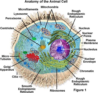
Diagram of animal cell under electron microscope

Diagram of a animal cell under light microscope
Animal cell structures
Characteristics;
- Have irregular shape.
- Have centrioles.
- Have lysosomes.
- Lack cell walls.
- Lack plastids.
- Store carbohydrates in the form of glycogen e.g. phagocytotic vacuoles, pinocytotic vacuoles, autophagic vacuoles and etc.
- Cytokinesis occurs by furrowing i.e. periphery – centres direction of constriction of cell membrane.
Structure of the plant cell
-
A plant cell is encased in a tough and rigid cellulose cell wall.
-
Beneath the cell wall is the cell surface membrane, which surrounds the cytoplasm.
-
The cytoplasm contains organelles, the prominent ones being:
- Vacuole
- Plastids (e.g., chloroplasts)
- Nucleus
-
Since a greater part of the cell is occupied by the vacuole, the cytoplasm and nucleus are squeezed to the periphery.
-
When viewed under a light microscope:
- Only a few structures are seen under high magnification
- Even finer details are seen at higher resolutions
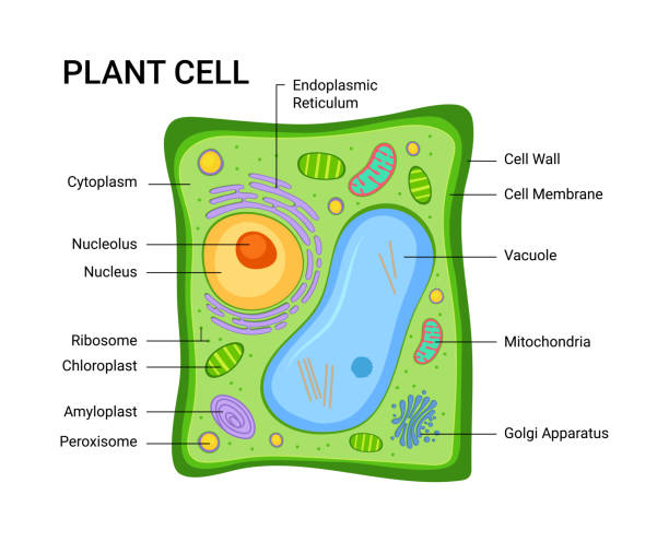
Diagram of a plant cell under light microscope

Characteristics of plant cells
- It has a fixed shape.
- It has a cell wall made up of cellulose.
- It has large permanent vacuole,
- It has plastids; chloroplasts, chromoplast and leucoplasts.
- Stores carbohydrates in the form of starch.
- Lack lysosomes.
- Lack centrioles.
- Cell division; cytokinesis follows centro-periphery direction.
Similarities between a plant and an animal cell
Both Have;
- Plasma membrane
- Distinct nucleus
- Ribosome
- Endoplasmic reticulum
- Cytoplasm
- Golgi apparatus
Qn What is an organelle?
An organelle is a distinct part of a cell which has a particular structure and function e.g. Mitochondria, chloroplast, ER etc.
Cell wall
Cell wall is the structure that occurs externally to the cell. Organisms with cell wall include.
- Bacteria – have cell wall made up of murein and peptidoglycan.
- Fungi – has cell wall made up of chitin.
- Algae and plant have cell wall made up of cellulose.
Plant cells cell walls
-
It is the structure external to the cell; it is not an organelle, although it is a product of various cell organelles
- E.g., microtubules and Golgi apparatus
Chemical composition
-
Made up of cellulose (mainly fibres) forming an amorphous matrix that surrounds the entire cell
- These fibres are made up of several hundred microfibrils, which form the network of the cell wall
-
In addition to cellulose, the plant cell wall also consists of:
- Pectin
- Hemicellulose
- These contribute to the mechanical strength of the organism
Pectin
-
These are polysaccharides of galactose and galacturonic acid
-
Pectin may combine with:
- Ca²⁺ to form calcium pectate
- Mg²⁺ to form magnesium pectate
-
These compounds are important components of the first layer of the cell wall to be laid down on the middle lamella
Hemicellulose
-
Hemicellulose is a mixture of many compounds, mainly:
- Sugars (e.g., glucose)
- Sugar acid residues
-
Hemicelluloses form hydrogen bonds with cellulose fibres in the cell matrix
-
The cell wall is usually modified by deposition of other substances such as:
- Alginic acid
- Calcium carbonate (in the case of algae)
Functions of cell wall
-
Mechanical support and skeletal support of individual cell and plants as well. This is through lignifications.
-
To prevent cell from bursting in hypotonic solution.
-
Control cell growth and shape. Orientation of cellulose microfibrils limits and helps to control cell growth and shape because of the cells ability to stretch is determined by their arrangements.
-
Movement of water and material salts. The system of interconnected cell walls (apoplast) is a major pathway of the movement of water and dissolved mineral salts. The cell walls are held together by middle lamellae, they also posses minute pores through which structures called plasmodesmata form living connections between cells and allows the protoplast to be linked in a system called symplast.
-
Reduction of water loss and reduced risk of infection (due to its waxy cuticle).
-
Transportation of materials. The walls of xylem vessels and sieve tubes are adopted for long transportation of materials through the cells.
-
Barrier to water movement. The cell walls of root endodermal cells are impregnated with suberin that forms a barrier to water movement.
-
Some cell walls are modified as food reserves as in the storage hemicelluloses in some seeds.
-
Transport of materials by active transport.
Cell organelles or organoids
-
Besides the cellular inclusions and plastids, the cytoplasm matrix contains many large-sized structures known as cell organelles or organoids.
-
These organelles perform various important functions such as:
- Synthesis
- Transportation
- Support
- Reproduction
-
Examples of organelles include:
- Endoplasmic reticulum
- Ribosome
- Golgi complex
- Lysosomes
- Mitochondria
- Plastids
- Centrioles
- Cilia
Functions of cytoplasm
- It provides medium for chemical reaction to take place like protein synthesis, lipids synthesis and etc.
- It stores useful materials such as amino acids, proteins, starch, carbohydrates, lipids,
- It stores waste materials such as and nitrogen waste etc.
- It controls the absorption of materials across the membrane due to its concentration gradient.
Endoplasmic reticulum (ER)
-
The cytoplasm matrix is traversed by a vast reticulum or network of interconnecting tubules and vesicles, known as the endoplasmic reticulum (ER).
-
The endoplasmic reticulum consists of a single vast and interconnected cavity bounded by a single membrane.
-
The ER membrane is believed to originate from pushings of the plasma membrane into the hyaloplasm (matrix), as it is chemically similar to a lipoproteinous structure like the plasma membrane.
-
The membrane of the ER can be:
- Smooth ER – when it does not have attached ribosomes
- Rough ER – when it has attached ribosomes
-
The membranes of the ER are continuous with both the nuclear membrane and the plasma membrane.
Functions of endoplasmic reticulum
- Transport of materials from exterior to the nucleus or to cytoplasm organelles such as Golgi complex.
- It provides mechanical support to the cytoplasm matrix.
- Functions as a cytoplasm framework. Surfaces for some of the biological activities of the cell catalyst its complex folding provide an enormous surface for such activities.
- Synthesis and transfer of lipids (smooth endoplasmic reticulum).
- In the liver the smooth endoplasmic reticulum detoxifies many poisons and drugs.
- The rough endoplasmic reticulum transports proteins synthesized in the ribosome of the rough endoplasmic reticulum.
- Formation of Golgi bodies as they are modified endoplasmic reticulum.
- Routes for movement of materials from the nucleus to the cytoplasm.
DIAGRAM 7

Golgi apparatus / Dictylosomes
This cell organelle is also known as the Golgi body, Golgi complex or sityasome.
DIAGRAM 8

It is the apparatus which consists of membranous sacs called cisternae and a system of small vesicle (called Golgi vesicles or dictysome vesicles) and vacuoles of various sizes. The membranes of Golgi complex are of lipoproteins and these are supposed to be originated from the membrane of endoplasmic reticulum.
Functions
Produce secretions
There are many Golgi apparatus in;
- Cells of salivary gland
- Cells of root cap
- Cells of endocrine glands i.e. pancreas
Modification of materials
The combination of carbohydrates and proteins to form glycoprotein takes place in them. Many materials such as mucin are glycoprotein. It takes place in the cistern. Carbohydrate chain + lipids = glycolipids
- Production of carbohydrates example cellulose produced in plants after division. Thus this separates one cell from another.
- Transport of lipids (storage and transport of proteins and lipids) after digestion, the fatty acids and glycerol are formed. In the endoplasmic reticulum fatty acids and glycerol unite to form lipids (triglycerides). The latter are passed to the Golgi apparatus where it transports them to the plasma membrane as lymphatic system and going to the lymphatic system.
- Formation of lysosomes.
- Synthesis of various types of carbohydrates from simple sugars.
- It activates the mitochondria to produce ATP.
- It forms the acrosome of the sperms.
Lysosomes
- Spherical, single membrane-bound organelles containing digestive enzymes.
- Enzymes are synthesized in ribosomes and rough endoplasmic reticulum (RER), then transported to the Golgi apparatus for modification.
- Golgi vesicles detach from the Golgi apparatus and remain in the cytoplasm as lysosomes because they contain digestive enzymes.
Functions of lysosomes
-
Function as storage vesicles for many powerful digestive (hydrolytic) enzymes.
-
Act as the digestive system of the cell, enabling it to process bulk materials taken in by phagocytosis or pinocytosis.
-
Digest parts of the cell such as:
- Worn out organelles
- Stored food contents of chloroplast A and B in extracellular digestion
-
Play a role in some developmental processes, e.g., remodeling of bones and fractures.
Note (NB)
- In plant cells, the large contractile vacuole may act as lysosomes.
- Bodies similar to lysosomes of an animal cell are sometimes seen in the cytoplasm of a plant cell.
Ribosomes (additional info)
-
Structurally composed of two subunits:
- Small subunit
- Large subunit
-
Each subunit is made of rRNA (ribosomal RNA) and proteins.
-
Present in both eukaryotic and prokaryotic cells.
-
Sizes determined by sedimentation during centrifuging, showing:
- 80S ribosomes (present in RER of eukaryotic cells)
- 70S ribosomes (present in prokaryotes and in mitochondria and chloroplasts of eukaryotic cells)
Adaptations of ribosomes
The ribosomes are the sites for protein synthesis. it has the following characteristics.
- Presence of enzymes capable of catalyzing the synthesis of peptide bonds.
- Presence of ribosomal RNA (rRNA) that attract other types of RNA i.e. mRNA and tRNA towards the ribosome's.
- Vacuoles
- A vacuole is a fluid filled sac which is bound by a single membrane.
Vacuoles
-
In animal cells, vacuoles are relatively small and temporary. Examples include:
- Phagocytotic vacuoles
- Pinocytotic vacuoles
- Autophagic vacuoles
-
In plant cells, the vacuole is large and occupies a greater proportion of the cytoplasm.
-
The membrane surrounding the vacuole is called the tonoplast.
-
The fluid inside the vacuole is called cell sap or vacuole sap.
Contents of cell sap
The cell sap is a mixture of many substances, including:
- Concentrated solutions of sugar and salt
- Organic acids
- Gases such as and
- Pigments
- Waste products of metabolism
It also contains enzymes similar to those found in lysosomes.
Roles of cell vacuoles
- They are involved in primary plant growth. It is a result of turgor pressure generated inside the vacuoles as a result of entry of water. This causes cell expansion as the tonoplast is pressed against the cell wall.
- The pigment contained in the cell sap is responsible for flower color and therefore play a key role to pollination.
- They contain enzymes similar to those of lysosomes when plant cell dies. The tonoplast looses the differential permeability and enzymes escape causing autolysis.
- Vacuole acts as a temporary store of waste products such as crystals of waste calcium oxalate, toxins and metabolic waste products of plants.
- The vacuoles sometimes functions as food reserves e.g. sucrose mineral salts and insulin are stored in vacuoles.
- In prokaryotes it serves for buoyancy.
Mitochondria
Structure of mitochondria
-
Shape: Sausage-shaped or oval-shaped organelle.
-
Surrounded by a double membrane called the mitochondrial envelope, which consists of:
- Outer membrane (smooth)
- Inner membrane (coiled to increase surface area for attachment of membranes)
-
Between the two membranes is the intermembranal space.
-
The inner membrane's coils increase the surface area for attachment.
Matrix (ground substance)
- Contains several enzymes responsible for the Krebs cycle.
- Contains circular DNA similar to that of prokaryotic cells, which allows for self-replication of mitochondria.
- Contains 70S ribosomes, like those in prokaryotic cells, responsible for protein synthesis (e.g., enzymes).
Diagram of mitochondrion

Functions of mitochondrion
-
The main function of mitochondrion is to yield energy during respiration.
-
About 98% of the cell's energy is synthesized in the mitochondrion.
-
For example, one molecule of glucose yields 38 ATP molecules.
-
Out of these 38 ATP, 36 ATP are synthesized in the mitochondrion through:
- Krebs cycle
- Electron transport chain
-
Because of this, mitochondrion is called the powerhouse, power station, or power plant of the cell.
Adaptations of the mitochondrion to energy production
- Presence of outer membrane and inner membrane to allow entry and exit of materials.
- The inner membrane is coiled to increase the surface area for attachment of enzymes responsible for electron transfer.
- Presence of matrix which is as granular and gives enough space for reaction to take place (Krebs cycle reaction) also matrix contains Krebs cycle enzymes.
- Presence of circular DNA for replication of the mitochondrion.
- Have 70s ribosomes for synthesis of proteins.
- Presence of phosphate for production of ATP.
- Presence of oxysomes and water accompany aerobic respiration.
Endosymbiotic theory (Evolution of mitochondria)
The mitochondria were originally independent prokaryotic bacteria like organisms which entered hosts cells and develop mutual relationship (symbiosis).
Mitochondria as prokaryotic cell
- Posses its own DNA and is able of self replication / reproduction.
- Have a circular like bacteria DNA.
- It is sensitive to different antibiotics such as chlorophyll and streptomycin which inhibit mitochondrial activities.
- It contains ribosomes similar to those of bacteria.
Plastids
These are organelles with double membrane, located in plant cells and algae. Types
- Chromoplasts
- Leucoplasts
- Chloroplasts
Chromoplasts
(Chromo – color / pigment) These are types of plastids bearing pigments i.e. yellow, red, orange, purple pigments. Found in
- Flowers
- Fruits
- Seeds
- Leaves
- Roots of carrots.
Leucoplast (embryos and germ cells)
Leuco- colour / white. These are colour plastids found mainly in storage organs. There are various types of leucoplasts;
- Amyloplasts- contain starch
- Lipoplasts – stores lipids
- Proteoplasts- stores proteins
Structure of chloroplasts
The chloroplast
The chloroplast is an oval shaped green in color due to presence of chlorophyll. It has two membranes an outer and an inner membrane which constitutes the double membrane or chloroplast envelope.

Functions of chloroplasts
-
It is the site of photosynthesis. This is the process whereby green plants manufacture food from and water in the presence of light energy, it stores starch temporarily.
-
The thylakoids have chlorophyll pigment for trapping sunlight energy.
-
It has grana and thylakoids to hold the chlorophyll in proper position for maximum absorption of light energy.
-
Stroma contains enzymes for dark reactions of photosynthesis.
-
Presence of phosphate which acts as a source of phosphate during phosphorylation.
-
Ribosomes and circular DNA for synthesis of proteins such as enzymes
Endosymbiotic nature of chloroplasts and mitochondria
-
Chloroplasts and mitochondria are endosymbiotic structures within a cell.
-
They can live independently inside a cell because:
- They have a double membrane which allows passage of materials in and out.
- They contain their own hereditary materials (circular DNA), enabling self-replication.
- They have 70S ribosomes that synthesize proteins (e.g., enzymes).
- They have a matrix or stroma, the ground substance where various reactions take place.
Details of structures
-
Stroma (in chloroplasts):
- Contains various photosynthetic membranes where light reactions occur.
- Dark reactions take place in the aqueous part of the stroma.
-
Matrix (in mitochondria):
- Site of the Krebs cycle of respiration.
-
Both organelles have their own enzyme systems.
Serial endosymbiotic theory
This theory accounts for the evolution of eukaryotic cells from prokaryotic cells. Evidence / similarities of organelle and prokaryotic cells
- Double membrane as cell membrane.
- Circular DNA.
- 70S ribosomes.
- System of enzymes.
Serial endosymbiotic theory (SET)
-
Suggests mitochondria and chloroplasts are descendants of ancient prokaryotic organisms.
-
Eukaryotic cells arose from the invasion of one large cell by other prokaryotic cells.
-
SET states:
- "All eukaryotic cells contain genetic material (DNA) and ribosomes that resemble those of prokaryotic cells."
-
Theory details:
- Prokaryotic heterotrophs ingested mitochondrion-like prokaryotes.
- At roughly the same time, an organized nucleus began to form.
- Non-motile cells established a symbiotic relationship with another prokaryote, such as spirochetes or spiroplasma bacteria attached to the outside of the cell, acting like a flagellum.
- Eventually, a photosynthetic prokaryote was engulfed, giving rise to a primitive plant cell.
Microbodies or peroxisomes
-
Small spherical bodies, 0.5 – 1.5 micrometers in diameter.
-
Ground substance contains important enzymes, especially catalase or peroxidase.
-
These enzymes catalyze the hydrolysis of hydrogen peroxide into water and oxygen.
-
Peroxisomes are found in:
- Liver
- Potatoes
- Pea seeds
- Bean seeds
Diagram
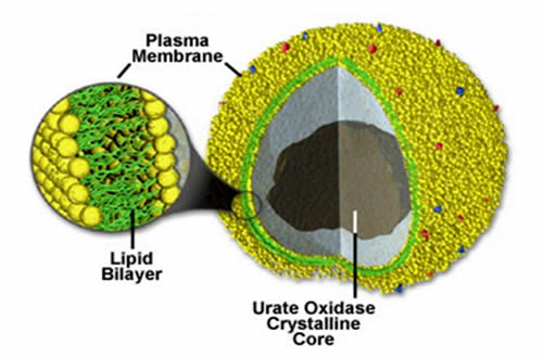
Functions of peroxisomes
- To break down the poisonous hydrogen peroxide to water and oxygen in the presence of peroxidase enzyme/ catalase.
- In plants special peroxisomes called glycoxisomes are centres for glyoxylate cycle i.e. conversion of fats into carbohydrates especially during germination.
Cytoskeleton
This is a complex network of fibrous protein structure that exists in cytoplasm of eukaryotic cell and anchor proteins or organelles such as nucleus to their fixed location.
The structures which constitute cytoskeleton include;
- Microfilament (actin filaments)
- Intermediate filaments
- Microtubules
Microfilaments (actin filaments)
These are thread like structures arranged in sheets or bundles first beneath the cell surface membrane. Chemically they contain actin and myosin. Each fibre is composed of two chains of protein loosely twisted about one another in helical manner. These proteins molecules can be assembled and dis-assembled.
Functions
- Interactions of these fibres with myosin help in muscle contraction. Determine the shape of cell's skeleton.
- Responsible for movement of materials within the cells.
- Cleavage of animal cells is brought about by the constriction of a ring of microfilaments after nuclear division, cytokinesis.
Intermediate filaments
These are structures intermediate between microtubule and microfilament (rope like microtubule of polypeptides) Skin cells for example form intermediate filaments from proteins called keratin. When the skin dies the intermediate filament of the cytoskeleton persists. Hair and nails are formed this way.
Function
- Provide cells shape
- Act as intercellular tendons preventing excessive stretching of cells.
Microtubules
- Microtubules are tubular structures made up of helically arranged globular subunit called tubulin.
- They are about 25 nm in diameter. Each has a chain of proteins wrapped round and round in a tight spiral. Large microtubules are found in cilia, flagella, centrioles (formation of spindle-fibres microtubules).
Functions
- They bring about movement of chromosomes during metaphase in nuclear division.
- Since they are tubular, they transport materials from one part of the cytoplasm to another, i.e. they are cytoconductors.
- In cilia and flagella, they help in rhythmical beating up movement.
- They determine the shape of the cell. (Skeletal support).
Cilia and flagella
The cells of many unicellular organisms and ciliated epithelium of multi-cellular organisms consists of some hair like cytoplasm projections outside the surface of the cell. These are known as cilia or flagella and they help in locomotion of the cells. The cilia and flagella are made up of proteins adenosine triphosphate (ATP). In prokaryotic cells, cilia and flagella (If they have structure lacking 9+2 arrangement of microtubules and arise from basal bodies). In eukaryotic cilia and flagella are complex. They have the 9+2 arrangement of microtubule and arise from basal bodies. Centrioles are present in animal cells only. They are two placed at right angle to each other. A number of rays called ultra rays usually surround the centrosomes. Each centriole is composed of nine paired thin threads and is in the form of cylinder. They aid in cell division.
Pinocytotic vesicle
These are organelle formed as a result of in folding of plasma membranes as it takes large particles of food from outside the cell. The process is called pinocytosis. Eventually pinch off and form very small vacuole (vesicle).
Functions
- Transport large particles into the cell.
- Nucleus is the functional unit of a cell.
- It contains materials which control different activities within the cell; the genetic materials.
Structure of the nucleus
The nucleus has a membrane called nuclear membrane envelope. Then nuclear membrane has some pores which allow some materials to pass in and out of nucleoplasm to allow communication on with cytoplasm called nuclear pores. Nuclear pores are made up of non-membrane materials forming nuclear pores. Nuclear envelope is semi permeable membrane allowing some materials to pass and others not to pass. The space inside the nucleus is filled by fluid materials which are called nucleoplasm. These are semisolid granules ground substance or matrix.

Within the nucleoplasm there are two components;
- Nucleolus
- Chromatin
- Matrix (aqueous)
Chromatin threads
Chromatin threads are grainy network of strands that undergo cooling into rod-like structures called chromatin. Chemically chromatin and therefore chromosomes contains DNA (deoxyribose nucleic acids) and much protein and some RNA (ribonucleic acids) and few minerals.
Nucleolus
These are small dark regions where different RNA type examples ribosomal RNA is produced and RNA joins the protein to form the subunit of ribosomes. It synthesizes the ribosomes protein and is used in controlling the cell division.
Functions of nucleolus
- Controls all metabolic activities of the cells
- It regulates cell division.
- Concerned with transmission of hereditary traits from parent to offspring.
- Synthesizes and stores proteins.
Mwalimu
Unasoma somo hili? Niulize nikuelezee chochote kilichomo.
Ingia ili kumuuliza Mwalimu wa AI wa Sonza kuhusu mada hii.
Ingia ili kuulizaMajadiliano
Hakuna maswali bado